← Atlas home All Views
Chapter 13

Memory and Learning

Brain structures involved in forming, storing, and retrieving memories. The memory circuit (hippocampus, mammillary bodies, thalamus, and cortex) is highlighted alongside the basal ganglia and cerebellum, which serve nondeclarative memory.
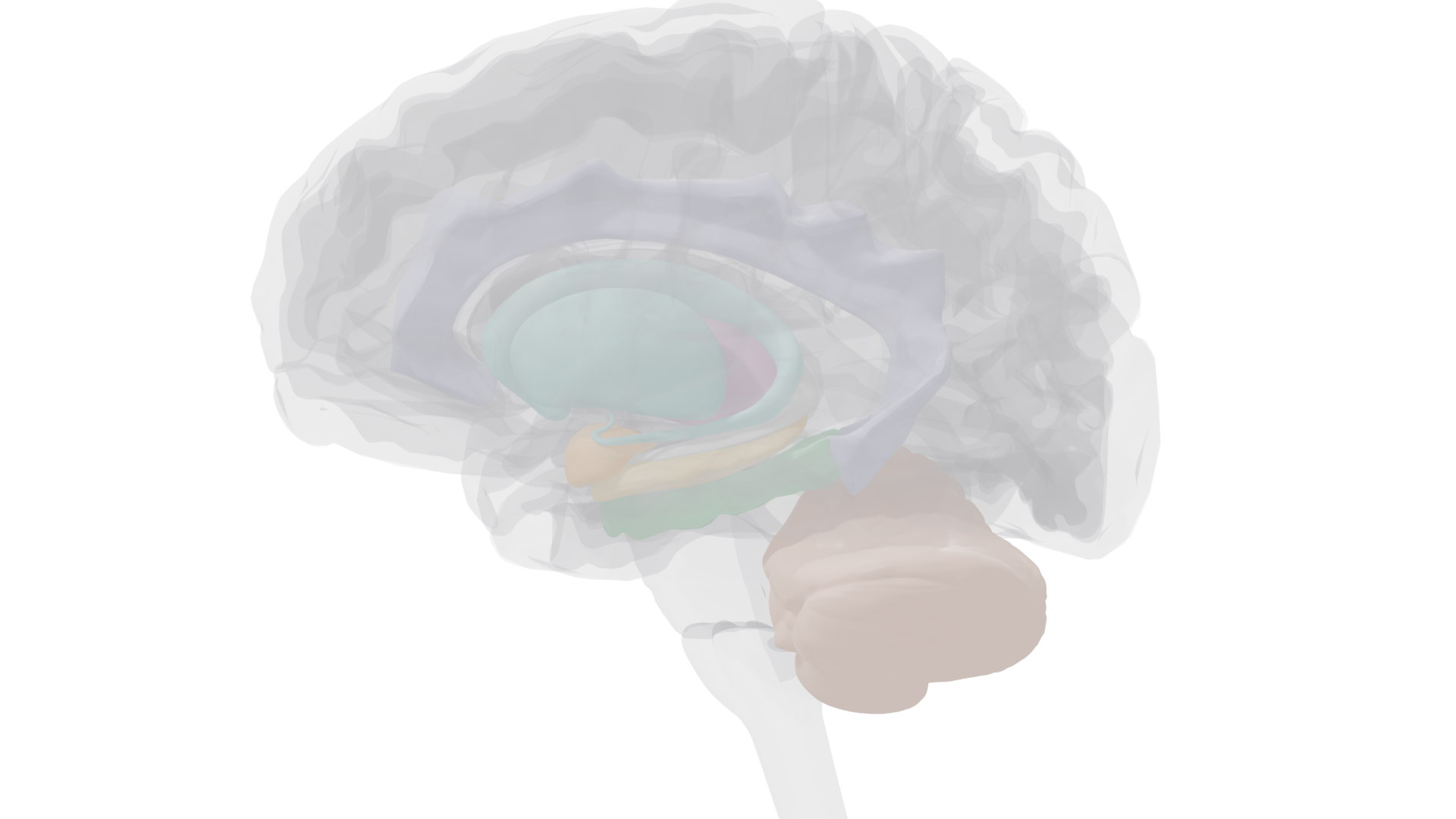
Midsagittal view highlighting hippocampus, thalamus, mammillary bodies, fornix, and cerebellum
Midsagittal (midline cut) view. Hippocampus (gold), thalamus (mauve), mammillary bodies (red), fornix (grey-white), and cerebellum (terra cotta) highlighted. Cortical structures ghosted.

Two Fundamental Types of Memory

Patient H.M.'s surgery revealed that memory is not a single system. He lost the ability to form new declarative memories but could still learn new motor skills. This dissociation defines the two major categories:

Declarative (Explicit)

Facts and events you can consciously recall and describe to others. Tested by asking "what" questions. Severely impaired in H.M.

Subtypes: Episodic (personal events tied to time and place) and Semantic (general knowledge without context of learning)

Hippocampus + Medial Temporal Lobe

Nondeclarative (Implicit/Procedural)

Skills, habits, conditioned responses, and priming effects shown by performance, not conscious recall. Tested by "how" tasks. Intact in H.M.

Subtypes: Skill learning, Priming, Classical conditioning, Operant conditioning

Basal Ganglia + Cerebellum + Cortex
Key distinction
Patient K.C. lost all episodic memory (couldn't recall any personal events) but retained semantic memory (general knowledge, chess skills). His cortical damage, not hippocampal, caused the selective episodic loss. This double dissociation (H.M. vs K.C.) shows episodic and semantic memory rely on partly different brain regions.

Landmark Cases

Patient H.M.: Henry Molaison (1926–2008)
Bilateral medial temporal lobectomy for epilepsy, 1953. Surgeon: William Scoville. Researcher: Brenda Milner.

Surgery removed most of the hippocampus, amygdala, and surrounding cortex from both temporal lobes. Result: profound anterograde amnesia, unable to form new declarative memories. Short-term memory was normal (could repeat a digit list), but information vanished as soon as he was distracted. Old memories from before surgery were mostly intact (retrograde amnesia was limited). Crucially, he could learn new motor skills (mirror tracing) without any memory of having practiced, proving that declarative and nondeclarative memory are separate systems.

Patient N.A.
Accident: miniature sword entered nostril, damaging medial diencephalon, 1960.

Damage to the dorsomedial thalamus and mammillary bodies, not the temporal lobe. Showed the same pattern as H.M.: normal short-term memory, intact nondeclarative memory, but severe anterograde amnesia for declarative information. This proved the hippocampus is part of a larger memory circuit that includes diencephalic structures.

Patient K.C.: Kent Cochrane (1951–2014)
Motorcycle accident causing extensive cortical damage plus bilateral hippocampal shrinkage.

Lost all episodic memory (no personal autobiographical recall) but retained semantic memory (general knowledge, conversation, chess). Could slowly acquire new semantic knowledge with spaced trials but never new episodic knowledge. The selective episodic loss was attributed to frontal and parietal cortex damage, not hippocampal damage alone.

The Declarative Memory Circuit

Three interconnected brain regions are required to form new declarative memories. Damage to any one produces anterograde amnesia. But established memories survive this damage; they are stored elsewhere, most likely in the cerebral cortex.

Hippocampus
Part of the medial temporal lobe; includes entorhinal, perirhinal, and parahippocampal cortices
The hippocampus is essential for consolidating short-term declarative memories into long-term storage. It contains place cells (neurons that fire when an animal occupies a specific location) and works with grid cells in the adjacent entorhinal cortex to form cognitive maps of space. Species that rely heavily on spatial memory (food-caching birds, polygynous voles with large home ranges) have proportionally larger hippocampi.
Key finding
Removing only the hippocampus impairs declarative memory; removing the hippocampus plus surrounding cortex (entorhinal, parahippocampal, perirhinal) produces even greater impairment. The severity scales with the extent of medial temporal lobe damage.
Mammillary Bodies
Paired structures on the ventral surface of the hypothalamus
The mammillary bodies connect the medial temporal lobe to the thalamus, serving as a relay in the declarative memory circuit. They are prominently damaged in Korsakoff's syndrome, a degenerative condition caused by thiamine (vitamin B1) deficiency, most commonly seen in chronic alcoholism.
Korsakoff's syndrome
Damage to mammillary bodies and dorsomedial thalamus produces amnesia plus two distinctive features not seen in other amnesias: confabulation (filling memory gaps with fabricated stories the patient believes are true) and anosognosia (denial that anything is wrong). These additional symptoms are attributed to concurrent frontal cortex damage. Cause: thiamine deficiency. Thiamine treatment can halt progression but cannot reverse existing damage.
Dorsomedial Thalamus
Medial diencephalon; relay between mammillary bodies and cortex
The dorsomedial thalamus receives input from the mammillary bodies and projects to the cortex. Damage here (as in Patient N.A.) produces the same pattern of anterograde amnesia as hippocampal damage, confirming it is part of the same memory circuit. Together with the mammillary bodies, it forms the medial diencephalic component of the declarative memory system.

Declarative Memory Formation Pathway

Information flows through a circuit from sensory input to long-term cortical storage. The hippocampal system is needed to consolidate memories, but not to store them permanently.
Sensory Input Encoding into STM
Hippocampus Consolidation
Mammillary Bodies Relay
Dorsomedial Thalamus Relay to cortex
Cerebral Cortex Long-term storage
Why H.M. kept old memories
Memories already consolidated in the cortex before surgery survived the removal of his hippocampus. The hippocampus is a gateway for forming new declarative memories, not a warehouse for storing them. This is why anterograde amnesia (can't form new memories) is more severe than retrograde amnesia (loss of old memories) in medial temporal lobe damage.
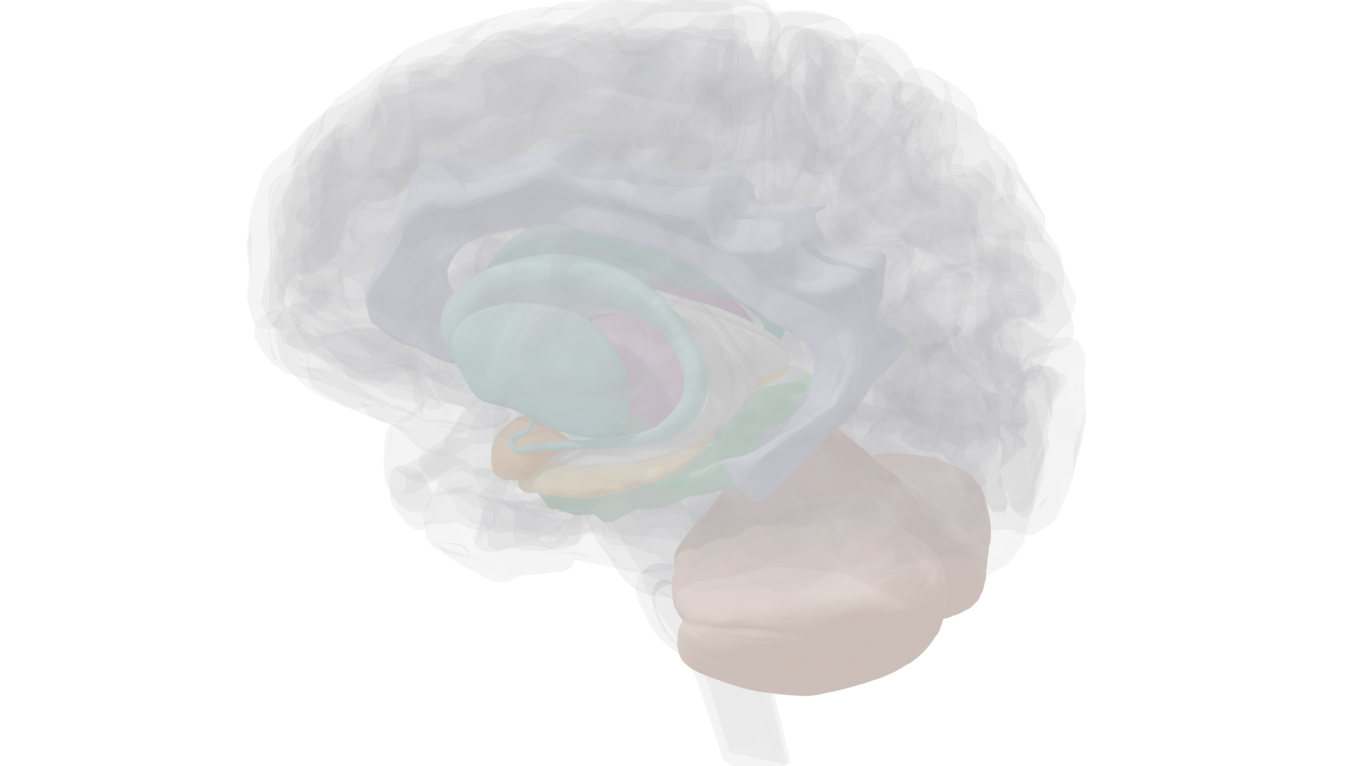
Deep structures view showing hippocampus, thalamus, basal ganglia
Deep structures (ghosted cortex): hippocampus, thalamus, basal ganglia, and cerebellum
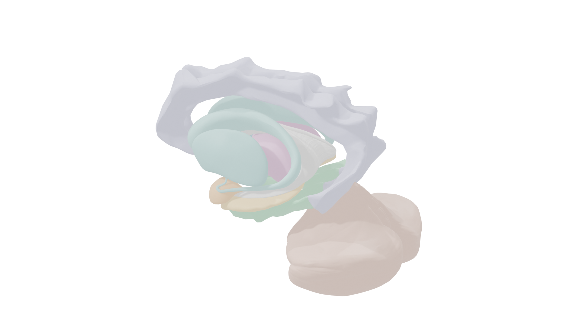
Isolated deep structures
Deep structures (isolated): memory circuit and nondeclarative memory regions

Nondeclarative Memory Systems

Different types of nondeclarative memory rely on different brain regions. None require the hippocampus, which is why H.M. could still learn skills.

Basal Ganglia
Caudate nucleus, putamen, globus pallidus
The basal ganglia are critical for skill learning: acquiring new sensorimotor, perceptual, and cognitive skills through deliberate practice. Damage to the basal ganglia impairs all three types of skill learning. The motor cortex and cerebellum also contribute to sensorimotor skill acquisition.
Types of skill learning
Sensorimotor (mirror tracing, riding a bike), Perceptual (reading mirror-reversed text), Cognitive (puzzle-solving like the Tower of Hanoi). All impaired by basal ganglia damage; sensorimotor also requires motor cortex and cerebellum.
Cerebellum
Crucial for classical conditioning of reflexes
The cerebellum is essential for classical conditioning, particularly the eye-blink reflex. Information about the unconditioned stimulus (air puff, via climbing fibers) and the conditioned stimulus (tone, via auditory pathways) converge on cerebellar neurons. After conditioning, the tone alone triggers an enhanced cerebellar response that produces the blink. Unilateral cerebellar damage eliminates conditioning on that side only.
Cortex
Occipitotemporal and left frontal regions
Priming, faster or biased processing of a stimulus because of prior exposure, is a cortical function. Perceptual priming (based on visual form) involves reduced activity in bilateral occipitotemporal cortex. Conceptual priming (based on meaning) involves reduced activation of left frontal cortex. Priming does not require declarative memory: amnesic patients show normal priming for items they cannot consciously recall.

Eye-Blink Conditioning Circuit

Classical conditioning of the eye-blink reflex illustrates how the cerebellum mediates associative learning. The CS and US converge in the cerebellum, where synaptic changes encode the learned association.

Air Puff (US) Cornea → Cranial nerve V
Brainstem Climbing fibers
Cerebellum CS + US converge
Tone (CS) Auditory pathways

Stages of Memory

Sensory Buffer Seconds
Short-Term Memory ~30 sec without rehearsal
Long-Term Memory Potentially permanent

Three processes move information through these stages: Encoding (sensory → STM), Consolidation (STM → LTM, requires the hippocampus for declarative memories), and Retrieval (LTM → working memory). Failure at any stage means information is lost.

Reconsolidation
Retrieving a memory from LTM makes it temporarily unstable before it is reconsolidated. During this window, memories can be distorted. Leading questions ("Did you see the broken headlight?" vs "Was the headlight broken?") can incorporate false details during reconsolidation. This is why repeated self-testing with feedback is one of the most effective study strategies, because each retrieval strengthens the memory through reconsolidation.
Emotional enhancement
Emotionally arousing events are remembered better because the amygdala enhances memory encoding in the cortex via adrenal stress hormones. Propranolol (a beta-blocker) eliminates this enhancement without changing the perceived emotional intensity of the experience.

Synaptic Plasticity & Long-Term Potentiation

Memory storage requires physical changes in the brain, specifically changes at synapses. The best-studied mechanism is long-term potentiation (LTP), a long-lasting increase in synaptic strength first discovered in the hippocampus.

LTP in the Hippocampus
Hebbian synapses: "cells that fire together wire together"
A brief high-frequency burst of stimulation (tetanus) to presynaptic neurons produces a long-lasting enhancement of the postsynaptic response. This depends on two glutamate receptor types working together:

AMPA receptors: mediate normal synaptic transmission. During low-level activity, these are the only active glutamate receptors.

NMDA receptors: blocked by Mg2+ during normal activity. Only become active when strong AMPA-mediated depolarization ejects the Mg2+ plug and glutamate is present. This dual requirement (depolarization + ligand) makes them coincidence detectors.

When NMDA receptors open, Ca2+ floods in, triggering enzymes that: (1) move additional AMPA receptors to the synapse, (2) increase existing AMPA receptor conductance, (3) stimulate production of new AMPA receptors, and (4) cause retrograde signaling that increases presynaptic glutamate release. The synapse is strengthened on both sides.

Evidence linking LTP to memory
Correlational: LTP time course matches memory formation. Blocking: NMDA antagonists impair LTP and spatial memory (Morris water maze). Knockout mice lacking hippocampal NMDA receptors can't form LTP or declarative memories. Enhancement: Mice overexpressing NMDA receptors show enhanced LTP and better long-term memory. Behavioral: Memory training induces measurable LTP in the hippocampus.

Environmental Enrichment & Brain Plasticity

Living in a complex environment with opportunities for learning produces measurable brain changes. Compared to animals in impoverished conditions, enriched-condition animals show:

• Heavier, thicker cortex (especially somatosensory and visual areas)
• Enhanced cholinergic activity throughout the cortex
• More dendritic branches and more dendritic spines on cortical neurons
• Larger cortical synapses
• More hippocampal neurons (newly generated neurons survive longer)
• Enhanced recovery from brain damage
Aplysia & simple learning
Even in the sea slug Aplysia, learning changes synapses. Short-term habituation: decreased neurotransmitter release at the sensory-motor synapse. Long-term habituation: actual reduction in the number of synapses. These findings from an animal with a simple nervous system reveal fundamental cellular principles that generalize across species.
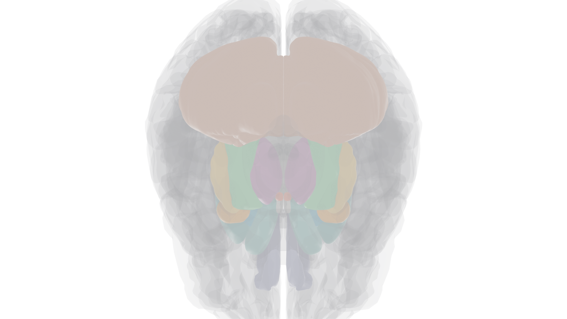
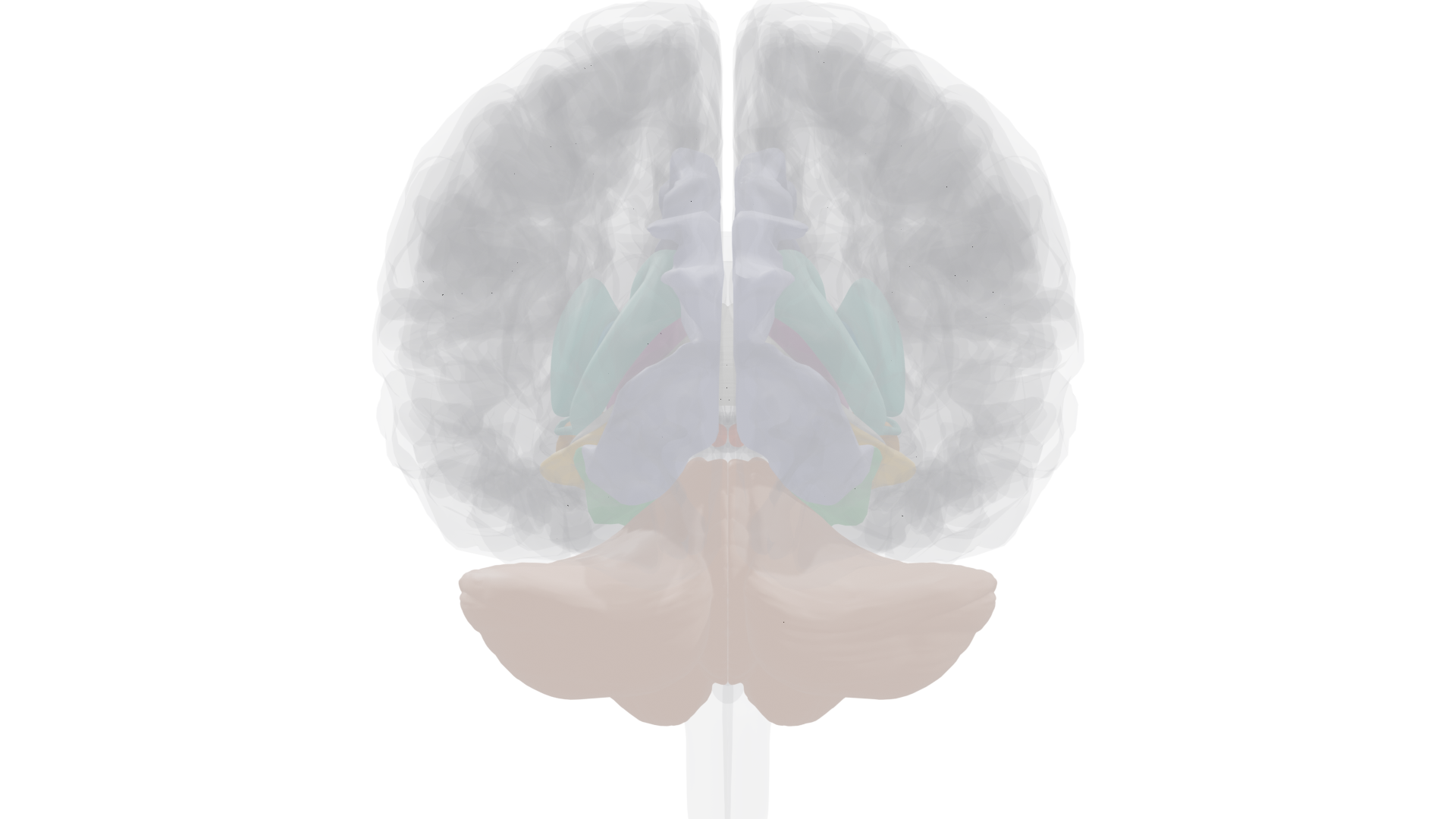
Anterior view showing thalamus and basal ganglia
Anterior (front): thalamus and basal ganglia visible through ghosted cortex
Ventral view showing hippocampus, mammillary bodies, and cerebellum
Ventral (bottom): hippocampus, mammillary bodies, and cerebellum from below

Key Terms

Anterograde amnesia
Inability to form new memories after brain damage or trauma
Retrograde amnesia
Loss of memories formed before brain damage (usually limited to hours or days)
Declarative memory
Explicit memory for facts (semantic) and events (episodic); requires hippocampus to form
Nondeclarative memory
Implicit memory shown by performance: skills, priming, conditioning; does not require hippocampus
Episodic memory
Autobiographical memories tied to a specific time and place
Semantic memory
General knowledge without memory of when or where it was learned
Consolidation
Process of converting short-term memory into long-term memory; requires hippocampus for declarative memories
Reconsolidation
Process by which retrieved memories become temporarily unstable before being restabilized; vulnerable to distortion
Place cells
Hippocampal neurons that fire when an animal is in or moving toward a specific location
Grid cells
Entorhinal cortex neurons that encode spatial position like a latitude/longitude grid
Long-term potentiation (LTP)
Long-lasting enhancement of synaptic transmission after high-frequency stimulation; candidate mechanism for memory
NMDA receptor
Glutamate receptor that acts as a coincidence detector; requires both depolarization and glutamate to open; critical for LTP
AMPA receptor
Glutamate receptor that mediates normal fast synaptic transmission; upregulated during LTP
Confabulation
Filling memory gaps with fabricated information that the patient believes is true; seen in Korsakoff's syndrome
Korsakoff's syndrome
Amnesia caused by thiamine deficiency (usually from alcoholism); damages mammillary bodies and dorsomedial thalamus
Cognitive map
Mental representation of the spatial layout of an environment; depends on the hippocampus
Engram
The physical memory trace: the neural change that encodes a specific memory
Priming
Changed processing of a stimulus due to prior exposure; does not require conscious recall